


All About Echocardiogram: An Overview

- Echocardiogram
- 22 Aug 2023
Overview
What is Echocardiogram ?
An echocardiogram, sometimes known as an “echo,” is a type of scan that may be used to examine the heart and the blood arteries that are close by. It’s a form of ultrasonic scan, which means a tiny probe is used to send out high-frequency sound waves that generate echoes when they bounce off different sections of the body. In other words, it’s a way to look inside people without cutting them open.1Overview| Researched based study from Nhs.uk
During the process of the scan, the probe will pick up on these echoes and convert them into a moving image that will be displayed on a monitor. Any doctor, including your primary care physician, who suspects that you may be experiencing issues with your heart may recommend that you get an echocardiography. A cardiologist is the medical term for a heart expert.
Uses
What purpose does an Echocardiogram serve?
During an echo test, your health care provider will be able to examine the anatomy of your heart as well as evaluate how effectively your heart is working.
Your health care team will be able to determine, with the aid of the test, the following aspects:
- The dimensions and form of your heart, as well as the measurements, thickness, and activity of the walls of your heart.
- The motions that your heart makes when it beats.
- The force with which the heart pumps blood.
- Assuming that the heart’s valves are functioning normally.
- If your heart valves are allowing blood to leak backwards, a condition known as regurgitation may be present.
- If there is an abnormal narrowing of the heart valves (Aortic stenosis).
- In the event that you have a tumor or an infectious development near your heart valves.2Uses| Researched based study from Heart.org
The test will also assist your health care provider determine if you have the following conditions:
- Issues with the pericardium, which is the protective layer that surrounds the heart.
- Issues affecting the big blood arteries that are responsible for entering and exiting the heart.
- The chambers of your heart get blocked with clots of blood.
- Abnormal openings in the walls of the heart’s chambers.
An echocardiogram examines the anatomy of the heart and the blood arteries that surround it, determines how blood flows through those blood vessels, and evaluates the chambers of the heart that are responsible for pumping blood throughout the body. Echocardiograms can be helpful in the diagnosis and monitoring of certain cardiac problems1Uses| Researched based study from Nhs.uk
Types
Types of Echocardiograms
- Transthoracic echocardiography (TTE)
- Transesophageal echocardiogram (TOE)
- Stress echocardiography
Transthoracic Echocardiography (TTE)
- The transthoracic echocardiography (TTE) is the type of echocardiogram that the majority of patients will get.3Types| Researched based study from Medlineplus.gov
- A piece of equipment known as a transducer will be positioned in a variety of ways on your chest and upper abdomen, and it will then be pointed in the direction of your heart. This apparatus generates sound waves of a very high frequency.
- The transducer is responsible for picking up the echoes that are left behind by sound waves and then transferring them into electrical impulses.
- These impulses are then transformed into moving photographs of the heart by the equipment that does echocardiogram. Additionally, still photographs are captured.
- There is also the possibility of three-dimensional images being shown. The section of the heart that is being analyzed as well as the type of equipment will determine the sort of image that is taken.
Transesophageal Echocardiogram
- A transesophageal echocardiogram, or TOE, is a procedure in which a small probe is passed down the throat into your food pipe (esophagus), and sometimes into your stomach (your throat will be numbed with a local anesthetic spray.
- You’ll be given a sedative to help you relax); you may need to refrain from eating for several hours prior to this test.
Stress Echocardiography
- Stress echocardiography is similar to a TTE; however, it is performed either during or immediately after a period of strenuous exercise, such as on a treadmill or exercise bike, or after an injection of a medicine that forces the heart to pump blood at a faster rate.3Types| Researched based study from Medlineplus.gov
- A harmless substance known as a contrast agent is injected into your circulation before an echocardiogram is carried out.
- This material shows up clearly on the scan and can help generate a better image of your heart. This is what is meant by the term “contrast echocardiogram.”
The Process
What takes place during the time of the Echocardiogram?
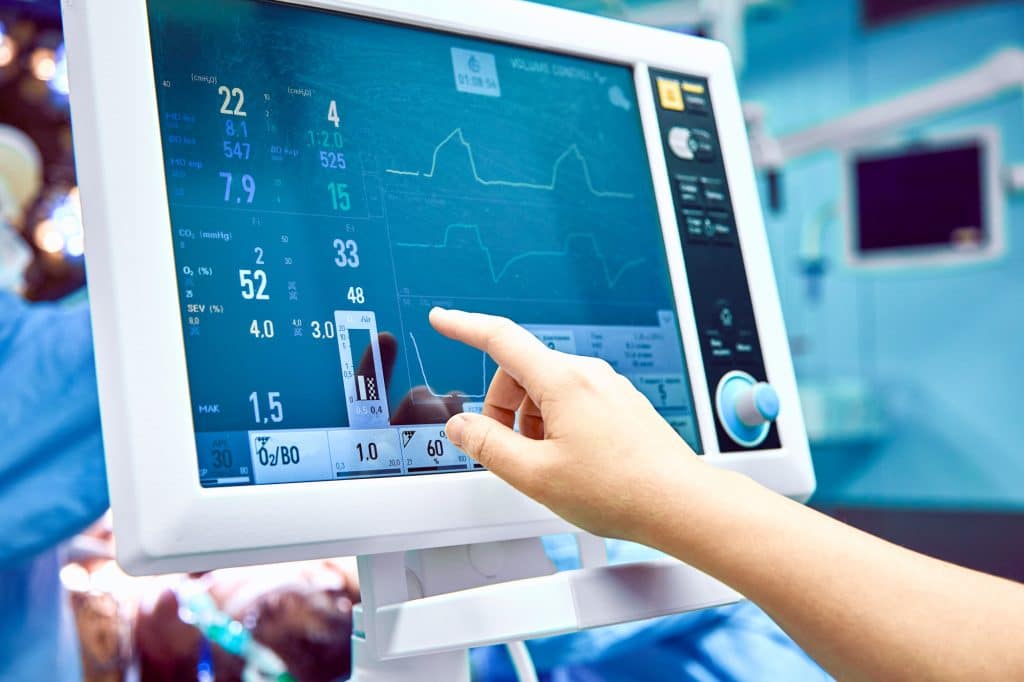
Echo exams are carried out by specialists who have received specialized training. Your doctor’s office, an emergency department, an operation room, a hospital clinic, or even your own hospital room are all potential locations for the performance of the test. The exam lasts for around one hour.
Steps are as follow:
- You will first be asked to lie down on a table, after which several thin metal disks, known as electrodes, will be attached to your chest. The disks are outfitted with cables that allow them to be attached to an electrocardiograph machine. During the exam, your heartbeat will be monitored using an electrocardiogram, often known as an ECG or EKG.2The process| Researched based study from Heart.org
- A gel is applied to your chest in order to facilitate the transmission of sound waves through your skin.
- In order to acquire clearer images, your technician may urge you to change positions or temporarily hold your breath.
- The probe (transducer) is moved over your chest in a back-and-forth motion. The probe generates sound waves, which then “echo” back to the probe after being reflected off of your heart.
- On a video monitor, the sound waves are converted into visuals after being processed on a computer.
- The images that are being displayed on the video monitor are being captured for your physician to review at a later time.
Side effects
Are there any potential adverse effects or risks of undergoing an Echocardiogram?
A normal echocardiography is a straightforward technique that does not cause any discomfort and is completely risk-free. The scan will not cause any adverse effects; however, the lubricating gel may feel chilly, and you may feel some little discomfort when the electrodes are withdrawn from your skin at the conclusion of the test.1Side effects| Researched based study from Nhs.uk ,4Side effects| Researched based study from Nhsinform.scot
- An echocardiography does not expose the patient to any radiation, in contrast to other diagnostic procedures and scans, such as X-rays and computerized tomography (CT) scans. On the other hand, certain of the less frequent varieties of echocardiograms come with a higher risk of complications.
- It is possible that you will have discomfort during the TOE operation, and subsequently, your throat may feel sore for a few hours. Because the sedative may cause you to feel sleepy for up to 24 hours following the exam, you will not be permitted to drive a vehicle during that time.
- There is also a remote possibility that the probe will cause damage to your throat.
- It is possible that you will feel queasy and lightheaded as well as have some chest discomfort while undergoing a stress echocardiography.
- In addition, there is a remote possibility that the process might cause an irregular heartbeat or a heart attack; nevertheless, you will be carefully watched throughout the test, and the operation will be terminated if any warning signals appear.
- During contrast echocardiography, the contrast agent that is administered to some patients causes them to develop an allergic response. In most situations, this will only produce moderate symptoms such as itching, but in extremely unusual circumstances, a severe allergic reaction may take place.
Precautions
What kinds of things should you avoid doing before getting an Echocardiogram?
It is dependent upon the sort of echo that you like to have done. Check with your service provider to find out exactly what you ought to stay away from. The following are examples of things you should try to avoid doing before your echo:5Precautions| Researched based study from Bhf.org.uk
- Eating or drinking.
- smoking cigarettes or utilizing any other goods containing nicotine.
- Consuming caffeine in any form, including coffee and other beverages. This includes caffeine-free beverages, some of which may have trace amounts of caffeine remaining. It also covers medicines available without a prescription that include caffeine in them.
- Before you go in for your echo, it is possible that your regular medication plan will need to be adjusted.
- Before discussing any modifications or stopping any drugs with your healthcare professional, you should not make any adjustments.
Takeaway
Takeaway
An echocardiogram, or echo, is a visual sketch of the motion of your heart. During an echo test, your healthcare professional will use ultrasonography, which is a form of high-frequency sound waves, to capture photos of the valves and chambers of your heart using a wand that is held in one hand and put on your chest. The healthcare professional can more accurately assess the pumping action of your heart with this information.
When assessing the flow of blood between the valves in your heart, medical professionals frequently mix the Doppler ultrasonography, echo, and color Doppler procedures.
There is no radiation involved in echocardiography. This distinguishes an echo from other diagnostic procedures such as X-rays and CT scans, both of which expose the patient to a little quantity of radiation.
Any feedback on this article?


 This Articles content was accurate
 Very Informative Article
 I have a question or a comment
 This article contains inaccurate content
 This article was not helpful
 I have a question or a comment
We appreciate your helpful feedback!
Checkout our social pages
References
-
National Health Service
Echocardiogram | Overview | Uses | Side effects
-
American Heart Association
Echocardiogram (Echo) | Uses | The Process
-
Medline Plus
Echocardiogram | Types
-
NHS Inform
Echocardiogram | Types | Side effects
-
British Heart Foundation
Echocardiogram | Precautions


Related Articles
-

What Are The Possible Causes Of Black Spot Inside The Mouth?
Introduction Black Spot Inside the Mouth It can be unsettling to discover a black spot or dot
-

Blood Cell Disorders : Types and Management
Overview What is Blood cell disorder ? Blood cell disorder is a term used to define any
-

Bowel Cancer Stomach Noises
Overview Bowel Cancer Stomach Noises Have you ever thought about what Bowel Cancer Stomach
-

Understanding Obesity: Causes and Management
Overview What is Obesity? Obesity is a complex medical disorder that causes adverse health
-

Shoulder Pain: Symptoms, Causes & Management
Introduction Shoulder Pain Any discomfort you experience near your shoulder joint is a