


Arrhythmia: Risk and Management

- Arrhythmia
- 14 Aug 2023
Overview
What is Arrhythmia ?
Arrhythmia denotes that the heart is not beating at a regular rate or rhythm. The heartbeat can be too slow, irregular, or too fast.
A normal resting heart rate in adults typically ranges from 60 to 100 beats per minute. Normally, heart rate will often increase when a person is physically active and decrease while at rest or sleeping. 1What is Arrhythmia? | Researched based study from nhlbi.gov It is also common to occasionally experience palpitations. But a persistent irregular heartbeat could indicate that the body is not receiving enough blood from the heart. 1.5 to 5% of the population may experience arrhythmias, the most frequent of which may be atrial fibrillation. 2What is Arrhythmia?| Researched based study from nih.gov Though arrhythmia is not typically significant, it can occasionally point to a cardiac condition that could be fatal.

Causes
Causes and risk factors
Arrhythmias may have a variety of causes or contributory factors, such as:
- Genetic factor – Arrhythmia can result from congenital heart problems and inherited conditions. It may run in families.
- Age – Chances increase with age since older people are more prone to develop various medical issues.
- Lifestyle – Tobacco, alcohol, and substance usage like opioids. 3Causes and risk factors | Researched based study from nih.gov
- Stress – Panic, surprise, or emotional stress disorders.
- Medicines – prescribed for various health issues can result in arrhythmia. For instance: Specific antibiotics, medicines for colds or allergies, etc.
- Heart problems – high blood pressure, coronary artery disease, cardiomyopathy, valve disorders, heart attack injuries, recovery from heart surgery, etc.
- Dehydration- electrolyte imbalances such as potassium or sodium in the blood may cause arrhythmia.
- Infections – viral illnesses such as flu or COVID-19. 4Causes and risk factors | Researched based study from nih.gov
- Other conditions – like diabetes, obesity, autoimmune disorders, sleep apnea, kidney diseases, lung disease, thyroid issue, etc. may also increase the chance.
- Environmental factors – like air pollution, 5Causes and risk factors | Researched based study from nih.gov can raise the risk of arrhythmia.
Classification
Types of arrhythmias
According to their place of origin, they can be divided into the following:
The supraventricular arrhythmias
It begins in the heart’s upper chambers and prevents blood from entering the heart’s chambers in between contractions. It may include:
- Premature atrial contractions – Extra beats that occur early and are not harmful.
- Atrial fibrillation – Most frequently appears in individuals over 65 years. 6Types of arrhythmia | Researched based study from nhlbi.gov Instead of creating a single, powerful contraction, the chamber fibrillates resulting in a heartbeat that is more than 400 times per minute.
- Atrial flutter – Those with heart problems and the first week following heart surgery are most likely to experience it. It typically develops as a result of one part of the atrium not conducting properly.
- Paroxysmal supra ventricular tachycardia (PSVT) – Generates additional heartbeats due to an issue with the electrical signals causing a fast heartbeat that typically has a regular rhythm. It starts and stops abruptly. It frequently affects young people and is typically not hazardous.
- Accessory pathway tachycardia – If there is a second pathway connecting the upper and lower chambers of the heart, this condition may induce a fast heartbeat.
- AV nodal reentrant tachycardia (AVNRT) – this irregular heartbeat is brought on by an additional channel through the AV node. Heart failure, fainting, or palpitations are all possible effects.
Ventricular arrhythmias
Starts in the lower chambers of the heart. These can be highly damaging and call for immediate medical attention.
- Premature ventricular contractions (PVCs) – are the most prevalent. These are the instances when we experience a skipped heartbeat.
- Ventricular tachycardia (VT) – a rapid, regular heartbeat that might last only a few seconds. More dangerous ventricular fibrillation may develop if this persists for a longer period than a few seconds. A past heart attack might leave a scar on the heart, which can lead to VT.
- Ventricular fibrillation – an irregular heartbeat that makes the bottom chambers tremble and unable to pump blood to the body. It frequently starts with a heart attack. Cardiac arrest and death can occur in a matter of minutes without the ventricles pumping blood to the body.
- Long QT syndrome – Too much time passes between contraction and relaxation of the heart’s bottom chambers. Death and life-threatening rhythm issues may result from this. It can be fatal and results in a rhythm called QT prolongation.
Bradyarrhythmia
Slow heartbeats can be caused by issues in the part of the heart’s conduction system. May consist of:
- An issue with the heart’s natural pacemaker results in sinus node dysfunction.
- Heart block- occurs when the path of the electrical impulse that the heart’s sinus node sends to its lower chambers is blocked.
Based on how they affect the heart rate, types are:
- Bradycardia: a resting heart rate of fewer than 60 beats per minute. 7Types of arrhythmia | Researched based study from nih.gov It occurs when the heart’s natural pacemaker stops functioning.
- Tachycardia: a resting heart rate of greater than 100 beats per minute while at rest.
- An early or extra heartbeat occurs when the heartbeat signal is sent too soon. When the heart resumes its regular rhythm, there is a pause followed by a harder beat.
Symptoms
Symptoms
There is a chance that they may not even produce any symptoms. The following symptoms if developed may include:
- Palpitations
- Dizziness
- Anxiety
- Fainting spells
- Pounding heart
- Chest pain
- Weakness or tiredness
- Breathing issues
- Gasping while sleeping
- A feeling of chest constriction
- Excessive sweating
- Cloudy vision
Prevention
Preventative measures
Arrhythmia is one of the heart conditions that cannot always be prevented. A person can lower their risk by doing the following things:
- Routine workout
- Reduce stress
- Keep hydrated
- Consume a balanced diet
- Steer clear of trans and saturated fats
- Keep blood cholesterol within normal range
- Maintain a healthy range of blood pressure
- Keep blood sugar under control
- Maintain a healthy weight
- Manage sleep apnea
- Stay away from tobacco use and smoking
- Reduce or stop drinking alcohol
- Cut back on or give up caffeine
- Get other medical issues treated
- To reduce the risk of sudden infant death syndrome (SIDS), 8Preventative measures | Researched based study from nichd.gov parents should adhere to safe sleep guidelines for newborns with an elevated risk of arrhythmia
Diagnosis
Diagnosis of Arrhythmia
The person will be questioned about their symptoms, family history, and lifestyle choices like drinking, smoking, etc.
Physical examination
- Check heart rate and rhythm, pulse, and blood pressure
- Looking for heart murmurs
- Checking for swelling in the legs or feet
- Looking for signs of other diseases like thyroid, lung disease
Blood test
- To evaluate potassium, electrolytes, and hormones in the blood
- Also, the presence of any infections could be detected
Electrocardiogram (EKG or ECG)
- It is frequently used to diagnose arrhythmias
- During a stress test, the doctor may perform an Electrocardiogram to capture the heart’s activity while it is working hard and beating quickly
Electrophysiology (EPS) test
- To monitor the electrical activity of the heart, catheters are implanted
- The optimal course of treatment for a specific patient is determined using the EPS to identify the reason for the aberrant rhythm
Cardiac catheterization
- This test helps identify whether coronary artery disease is the source of the arrhythmia
- Also offers details on the functionality of the heart’s muscles and valves
Holter monitor
- It is a portable electrocardiogram (EKG), often known as an ambulatory electrocardiogram or an ECG, that is roughly the size of a postcard or digital camera and that a person can use for up to two weeks.
- The heart’s electrical activity is measured during the test.
Event monitor
- The doctor might advise the patient to wear one of them, typically for about a month, if the symptoms do not occur frequently
- When a button is pressed, the device records and temporarily saves the electrical activity of the heart. When symptoms appear, attempt to get a reading. The results will be interpreted by the physician
Implantable loop recorder
- The doctor inserts this tiny gadget just beneath the skin of the patient, where it continuously logs the electrical activity of the heart
- The doctor’s office may receive information from it
Stress test
- The objective is to determine the maximum amount of stress that the heart can withstand without developing a rhythmic issue or not receiving enough blood.
- The patient will walk on a treadmill or ride a stationary bike during the most frequent type of stress test, while also getting an EKG, their heart rate and blood pressure are monitored, and they will also have an EKG taken
Tilt table testing/passive head-up tilt test/head upright tilt test
- When receiving an EKG and having blood pressure and oxygen saturation levels checked by medical professionals, the patient will need to lie on a stretcher at various angles.
- This identifies if the patient’s electrical, neurological, or vascular systems are to blame for their fainting symptoms.
Genetic testing
- It can determine if a person suffers from an arrhythmia that is triggered by a particular gene.
- This test is crucial if one or more family members have experienced a cardiac arrest or an arrhythmia brought on by the same gene.
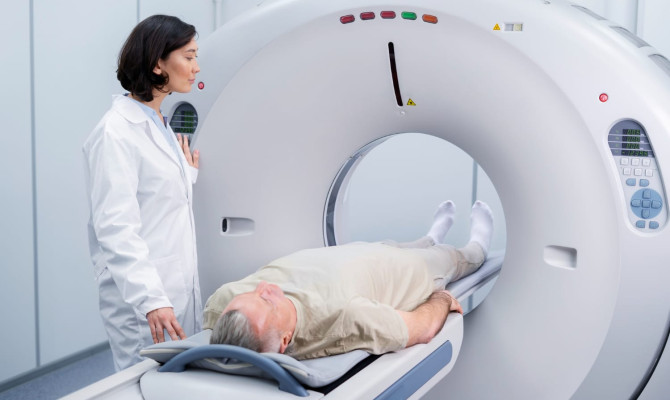
Heart imaging tests
- Such as cardiac computed tomography (CT) scans or MRIs, which can check the anatomy of the heart chambers, demonstrate how well the heart is functioning, search for signs of scar tissue in the heart muscle, and occasionally even look at the heart arteries.
Echocardiogram
- An ultrasound technique that gives a glimpse of the heart and helps doctors assess whether a patient’s heart valve or muscle dysfunction may be the source of an arrhythmia.
Complications
Complications associated with Arrhythmia
Without therapy, an irregular heartbeat could result in harmful issues like:
- Heart failure – Heart failure can happen as a result of persistent bradycardia or tachycardia. When the heart stops working, it is unable to provide the body’s organs with adequate blood.
- Stroke – The heart’s pumping efficiency is compromised by A-fib. Blood may gather in pools as a result and clot. If a clot moves, it might go to the brain artery and block, which could be fatal, or it might result in a stroke.
- A heart attack – occurs when blood flow is cut off to a section of the heart muscle. Heart cells will start to die if they are not given oxygen.
- Cognitive disorders – Dementia and Alzheimer’s disease may develop over time as a result of inadequate blood flow to the brain.
Treatment
Treatment options for Arrhythmia
Arrhythmia’s severity and kind will affect the course of treatment. If there is an underlying ailment, there is a risk of complications, or there are severe symptoms, an arrhythmia patient may require therapy. Options for treatment include:
Medicines
Arrhythmias can be treated with a wide range of medications. Sometimes medications are used with other medical procedures.
- Anti-arrhythmic medications can either be used to treat arrhythmias or prevent them from occurring.
- A fast heartbeat can be treated with certain medicines like beta-blockers, calcium channel blockers, digoxin, adenosine, etc.
- Medicines that treat uneven heart rhythms may be used.
- Drugs that lessen the chance of blood clots or strokes include aspirin and warfarin, both of which are blood thinners.
- Related disorders that might be producing an irregular cardiac rhythm can also be treated with medications.
Vagal maneuvers
- By activating the vagus nerve, which aids in controlling your heart rate, your body is made to relax.
- The doctor may instruct the patient to cough or spit, hold their breath, lie down, or cover the face with a cool, damp towel.
Invasive therapies may include
Electrical cardioversion:
- Drug therapy alone might not be enough to restore a normal rhythm in patients with enduring previous or irregular arrhythmias.
- In these situations, the doctor does cardioversion.
Catheter ablation:
- The little patch of heart tissue that is responsible for the abnormal heart rhythm is given high-frequency electrical energy via a catheter.
- The irregular rhythm’s route is disconnected by this energy.
- Atrial flutter, the majority of SVTs, and some ventricular and atrial tachycardias can all be treated with ablation.
Pulmonary vein isolation:
- it is a form of ablation that targets regions suspected to be the origin of atrial fibrillation in individuals with frequent atrial fibrillation.
- The intention is to produce scar rings that isolate the atrial fibrillation-causing foci.
Treatment with devices
During an operation in the electrophysiology lab, a cardiologist may insert particular devices.
Permanent pacemaker:
- It is a device that electrically stimulates the heart muscle with tiny pulses to keep the heart rate safe.
- To prevent your heart from beating too slowly, pacemakers are typically used.
An implantable cardioverter-defibrillator (ICD):
- It is an advanced medical gadget used primarily to treat life-threatening heart arrhythmias like ventricular tachycardia and ventricular fibrillation.
- The ICD tracks the cardiac rhythm continuously and when it notices an extremely rapid, abnormal heartbeat, it sends energy to the heart muscle to help the heart resume beating normally.
Biventricular (B-V) pacemakers / cardiac resynchronization therapy (CRT):
- Used in patients with uncoordinated contraction in addition to heart failure.
- These tools facilitate the left ventricle’s synchronized contraction.
Surgery
Surgical treatment
A person may require cardiac surgery to correct a heart condition that could be the cause of the arrhythmia, such as a valve or coronary artery bypass surgery:
- Atrial fibrillation can be treated surgically with the Maze and modified Maze procedures. The heart’s upper chambers are cut through by the surgeon. The intention is to confine the electrical impulses of the heart to specific routes.
- Some individuals could require a pacemaker thereafter.
- When additional heart procedures, such as valve surgery, are intended, it is frequently used.
- In some circumstances, endoscopic or minimally invasive procedures may be used to implant biventricular pacemaker leads in the heart.
Takeaway
Key Takeaways
Arrhythmias are frequently not harmful, but they can indicate a more dangerous condition. This makes it crucial to seek medical help if someone experiences arrhythmia symptoms. Arrhythmias cannot always be avoided, but frequent exercise, eating a nutritious diet, and getting treated for underlying disorders can all help. A cardiologist should be consulted if someone exhibits any of the symptoms. They may determine the existence of an arrhythmia, identify its causes, and begin developing a treatment plan.
Any feedback on this article?


 This Articles content was accurate
 Very Informative Article
 I have a question or a comment
 This article contains inaccurate content
 This article was not helpful
 I have a question or a comment
We appreciate your helpful feedback!
Checkout our social pages
References
-
National Heart, Lung, and Blood Institute
What Is an Arrhythmia? | Overview
-
National Institutes of Health
Clinical Epidemiology of Atrial Fibrillation and Related Cerebrovascular Events in the United States | Overview
-
National Institutes of Health
Opioids and Cardiac Arrhythmia: A Literature Review | Causes
-
National Institutes of Health
Arrhythmia in COVID-19 | Causes
-
National Institutes of Health
Air pollution and incidence of cardiac arrhythmia | Causes
-
National Heart, Lung, and Blood Institute
ATRIAL FIBRILLATION Causes and Risk Factors | Types
-
National Institutes of Health
Sinus Bradycardia | Types
-
Eunice Kennedy Shriver National Institute of Child Health and Human Development
How can I reduce the risk of SIDS? | Preventative measures


Related Articles
-

Understanding Stomach Cancer: Causes, Symptoms, and Treatment
Overview What is Stomach Cancer? Stomach cancer, also known as gastric cancer, starts when
-

What is Osteoarthritis: Symptoms and Treatment
Overview What is Osteoarthritis? Osteoarthritis or OA, is the most common type of arthritis.
-

Diagnosis & Management Of Tongue Tie In Adults
Overview Tongue Tie Ankyloglossia, is also known as tongue-tie. It is a condition that limits
-
Everything You Need To Know About Gout In Heel
Overview Gout Gout is a kind of arthritis that usually affects the joint at the big toe base
-

Turtle Neck Syndrome: Symptoms, Causes, Prevention & Treatment
Introduction Turtle Neck Syndrome A strange health condition known as Turtle Neck Syndrome